What Are the Benefits of Surgical Robots?
Follow article
 Dave from DesignSpark
How do you feel about this article? Help us to provide better content for you.
Dave from DesignSpark
Thank you! Your feedback has been received.
Dave from DesignSpark
There was a problem submitting your feedback, please try again later.
Dave from DesignSpark
What do you think of this article?
Surgeons have always relied on specialized equipment during patient procedures, with their options getting more advanced through the generations. Many hospitals now feature surgical robots to assist with operations. They don’t replace surgeons but instead provide numerous benefits to specialists and support staff.
Offering More Comfort and Precision for Surgeons
The first surgical robot arrived in 1985. Surgeons used it to insert a needle into a patient’s brain to take a biopsy. Before the availability of robots, that procedure had a high error rate due to human hand tremors. Robotic controls don’t have that shakiness. That’s one of the main reasons why surgeons like using them for delicate procedures that could result in severe complications if problems arise.
When surgeons use robots, they use controllers to guide their movements. The most common hand placements used when doing this are the pinch grip and the power grip. When performing a pinch grip, a person uses their thumb and first two fingers. Conversely, the power grip involves grasping something with the whole hand.
Pinch grips offer the most precision, but they’re uncomfortable to sustain for long periods. Power grips do not support detail-oriented movements and are better for tasks that require great force. They’re more comfortable, however.
A team of Japanese researchers engineered a surgical robot controller that allows using both kinds of grips. A proof-of-concept experiment showed that users preferred the new design, praising it for its comfort, stability, and ease-of-use.
Allowing More Collaborative Procedures
Many surgical robots take up substantial space. Their size often makes it difficult for surgeons to quickly intervene when needed.
To answer this challenge, a new model called Dexter boasts a better and more streamlined design that puts the surgical specialists closer to their patients. It allows switching between manual laparoscopic procedures and those performed by robots.
German surgeon Georg Kelling performed the first laparoscopic procedure in 1901. Other medical professionals quickly noticed their benefits, such as smaller incisions that shortened patients’ recovery times.
However, traditional methods for performing this surgery require standing for hours. This model offers ergonomic improvements that enable surgeons to move between sitting and standing positions throughout operations. They also stay close to patients while the robot moves.
This robot permits more collaboration between humans and machines. Design director Marcus Heneen explained:
“We know from experience that human-robot interaction works best when technology caters to the social setting in which it is deployed. Surgery is not a purely mechanical task of making incisions and joining tissue. It’s a collaborative process between surgeons and their teams, with a strong social component to it.”
Facilitating Remote Operations
Surgical robots often have tungsten cables and assemblies that allow the movement of the arms and other attachments that perform procedures. All of these machines require stringent testing to ensure they’ll work as expected during lifesaving operations.
One common test involves using a special device to proof load cables to 60% of their minimum holding strength. This evaluation ensures that crucial components can withstand applied tension without failing.
Testing is arguably even more important considering the rise of remote operations where the surgeon may be miles away from a patient.
A system called CorPath GRX is a cable-facilitated robotic system for performing percutaneous coronary interventions. These procedures involve inserting stents to open blood vessels in the heart that become narrowed due to plaque buildup. A 2019 research paper described how surgeons could use the machinery to perform procedures while 20 miles away from the patient.
Five people consented to this type of approach. All had the percutaneous coronary interventions done successfully and did not experience any major adverse cardiac events before discharge. There was a team onsite in each case to step in if necessary.
However, the remotely located surgeon did everything required with no need for the team’s help. This achievement could prove especially significant for people who live in areas that lack enough medical specialists at local hospitals to bear the patient caseloads.
Moving Ahead With Microrobots
The earlier example of Dexter shows that surgical robots are getting much smaller than the earliest models. That’s progress, but likely not the extent of what’s possible. Researchers recently used an origami-inspired technique to create a comparatively tiny bot. It’s about the size of a tennis ball and weighs as much as a penny. It may be a while before you see widespread usage of these designs, but the possibilities they offer are fascinating.
The team built the creation with a manufacturing technique called Pop-Up MEMS. It involves materials deposited on top of each other in bonded layers. These are then laser-cut into a precise pattern that allows them to rise in a 3D shape, similar to the illustrations in a children’s pop-up book. This technique facilitates mass-producing small and complex structures that would otherwise take much longer to do by hand.
Testing for the bot occurred via a mock retinal vein cannulation procedure. Surgeons perform it by inserting a needle into the eyeball to deliver therapeutics to the veins in the back of that body part. A retinal vein is about twice the thickness of a human hair strand. The team created a silicone tube of that diameter to check the robot’s accuracy. The bot punctured it with a needle and did not cause any damage or disruption to the surrounding area.
Reaching Deeper, Harder-to-Access Places
Whether surgeons perform traditional or robotic surgeries, they face continual challenges related to safely accessing some of the most out-of-reach places in the body.
A recent advancement may hold valuable answers. It’s a robotic platform that relies on the fringe field generated by an MRI scanner's superconducting magnet. Its force guides medical instruments through deep blood vessels.
The natural friction inside blood vessels combined with the ultra-small tools surgeons use when working with vascular structures create scenarios where the instruments can get stuck as surgeons try to move them to the right places.
However, this recent experiment involved placing a magnet on an instrument’s tip, then using the MRI scanner’s fringe field as an outside attracting force that pulled the tool along inside the body.
So where did robots come into the picture?
The team came up with a robotic table that sits inside the MRI fringe field. A patient lies on it, then automatically gets oriented in the right position relative to the instrument's current position in their body. A technique called fringe field navigation maps the directional forces of the MRI scanner’s magnetic field. That data then triggers the table to move the patient to the best orientation.
Surgical Robots on the Rise
These examples give a fascinating glimpse into the progress associated with surgical robots. They’re not the best choices in every case, but they often provide notable advantages over more familiar types of surgeries.
Surgical professionals’ overarching goals are to perform the necessary procedures successfully and safely while investigating methods that improve patient outcomes and reduce complications. Robots could help them succeed in those aims.
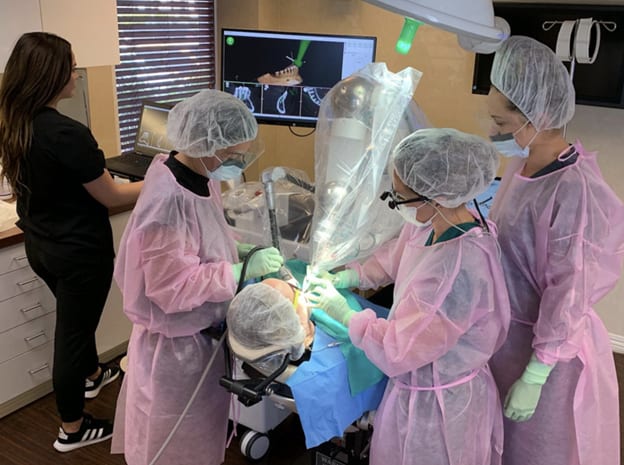
Featured image credit: Neocis Inc., CC BY-SA 4.0, via Wikimedia Commons